 ultimate-guide
Causes of Peptide Experimental Failure: 2026 Guide
Table of Contents
- Why Peptide Experiments Fail: An Overview
- Signs of Peptide Degradation Every Researcher Should Know
- Peptide Aggregation Prevention: Strategies for Reliable Assays
- Best Practices for Peptide Storage to Avoid Failure
- Analytical Validation Techniques for Peptide Quality
- Standardized Troubleshooting Workflow for Peptide Experiments
- Conclusion
- Frequently Asked Questions
Last Updated: September 15, 2026
Why Peptide Experiments Fail: An Overview
The causes of peptide experimental failure rarely come down to a single dramatic mistake. More often, it's a slow accumulation of small variables, each one nudging your results off course until the assay simply doesn't work. This guide from BlueWaveCompound breaks down the most common causes of peptide experimental failure and gives you a practical framework for diagnosing them.
At its core, peptide experimental failure is the inability of a peptide reagent or assay system to produce reproducible, interpretable results under controlled conditions. It typically stems from degradation, aggregation, or handling errors rather than from the underlying biology.
The tricky part is that these failures often look identical on the surface. A flat standard curve could mean your peptide degraded, aggregated, or was never fully soluble to begin with. Below, we'll walk through the specific failure modes, how to spot them, and what to do about them.
Signs of Peptide Degradation Every Researcher Should Know
The clearest signs of peptide degradation are a drifting standard curve, declining signal over repeated runs, and visible changes in the physical appearance of the powder or solution. If your assay worked last month and fails now with the same reagent, degradation is the first suspect.
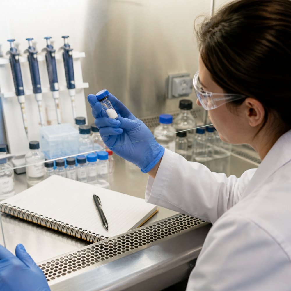
Watch for these specific indicators:
- Cloudiness or precipitate in a solution that was previously clear
- Color shift in lyophilized powder, from white to yellow or brown
- Reduced peak height on HPLC compared to your reference chromatogram
- New peaks appearing that weren't in the original analysis
- Inconsistent bioactivity across replicates from the same vial
Chemical degradation factors like pH, light, and heat drive most of these changes. Peptides with oxidizable residues such as methionine, cysteine, or tryptophan are especially vulnerable to light and oxygen exposure.
A common mistake is assuming a sealed vial is a protected vial. It isn't. Lyophilized peptides can still absorb moisture from the air each time the vial is opened, and that moisture accelerates hydrolysis.
Peptide Aggregation Prevention: Strategies for Reliable Assays
Peptide aggregation prevention starts with understanding your sequence's hydrophobicity. The more hydrophobic the sequence, the higher the aggregation risk, and the more care you need during solubilization and storage.
Aggregation happens when peptide molecules self-associate into dimers, oligomers, or larger fibrils. These aggregates often retain biological activity but with altered pharmacokinetics and unpredictable assay behavior. They can also form secondary structure that changes how the peptide interacts with your target.
Strategies that work in practice:
- Solubilize in small increments rather than dumping powder into a large volume at once
- Use a chaotropic agent like guanidine hydrochloride if the peptide resists dissolution
- Add a low percentage of organic solvent such as acetonitrile for hydrophobic sequences
- Avoid vortexing aggressively, which introduces shear that promotes aggregation
- Filter through a 0.22 µm filter before running critical assays
If you're working with a sequence that aggregates no matter what you try, consider whether your buffer composition is working against you. High salt concentrations and pH near the peptide's isoelectric point both increase aggregation tendency.
Best Practices for Peptide Storage to Avoid Failure
Best practices for peptide storage come down to three variables: temperature, moisture, and light. Control all three and you'll extend the useful life of your peptide considerably.
Store lyophilized peptides at -20°C for short-term use and -80°C for long-term storage (Recommendations for the generation, quantification, storage and handling of peptides used...). Desiccate the vial before sealing if you're storing for more than a few months. Protect from light with amber vials or foil wrap, particularly for sequences containing aromatic residues.
| Storage Condition | Recommended Temperature | Typical Duration | Key Risk |
|---|---|---|---|
| Lyophilized, sealed | -20°C | 6-12 months | Moisture ingress |
| Lyophilized, long-term | -80°C | 1-2 years | Freeze-thaw cycling |
| Reconstituted, working | 4°C | 1-2 weeks | Microbial growth |
| Reconstituted, archived | -80°C | 3-6 months | Aggregation on thaw |
Reconstituted peptides are far less stable than powder. If you need to keep a working solution for more than a week, aliquot it and freeze at -80°C. Never refreeze a thawed aliquot.
Analytical Validation Techniques for Peptide Quality
Analytical validation is what separates a reproducible experiment from a guessing game. Before you trust any peptide in a critical assay, confirm its identity, purity, and structural integrity with independent methods. Most practitioners find that running at least two orthogonal techniques catches the errors a single method misses.
Reverse-phase HPLC (RP-HPLC) remains the standard for purity assessment. It separates peptides by hydrophobicity and reveals truncation products, oxidation, and deletion sequences as separate peaks. A typical run uses a C18 column, a water/acetonitrile gradient with 0.1% trifluoroacetic acid (TFA) as the ion-pairing agent, and UV detection at 214 nm (peptide bond) or 280 nm (aromatic residues). A single sharp peak at the expected retention time is a good sign, but it does not confirm identity. Watch for shoulder peaks, which often indicate deamidation or diastereomer formation.
Mass spectrometry (ESI-MS or MALDI-TOF) confirms molecular weight and catches sequence errors that HPLC can miss. Compare the observed mass to the theoretical monoisotopic mass calculated from the sequence. A peptide that is 99% pure by HPLC but has the wrong mass is still the wrong peptide. For sequences under about 5 kDa, ESI-MS typically resolves the charge envelope cleanly; for larger or hydrophobic peptides, MALDI-TOF with a sinapinic acid matrix is often more forgiving (pubmed.ncbi.nlm.nih.gov).
Circular dichroism (CD) spectroscopy is the underused third check. It reports on secondary structure, alpha-helix, beta-sheet, random coil, and can flag a peptide that has folded or aggregated in a way that HPLC and MS cannot see. A far-UV scan from 190 to 260 nm takes minutes and is especially valuable for peptides intended for structural or binding assays.
Amino acid analysis (AAA) remains the reference method for compositional accuracy. It will not detect sequence scrambling, but it will catch a peptide that is missing an entire residue or has the wrong ratio of a key amino acid.
For reproducibility across batches, request a certificate of analysis from your supplier and check that the reported purity, mass, and appearance match your own in-house testing. At BlueWaveCompound, every batch ships with third-party lab testing and a certificate of analysis, which takes the guesswork out of this step. You can review the documentation standards at https://bluewavecompounds.com.
A practical acceptance checklist before committing a batch to a critical assay:
- Purity by RP-HPLC at or above the supplier's stated specification, with no unexplained shoulder peaks
- Observed mass within the method's tolerance of the theoretical mass
- CD spectrum consistent with the intended conformation
- Appearance and solubility match the certificate of analysis
- No new peaks compared to the reference chromatogram from the previous batch
FDA guidance on analytical procedures and methods validation
If any of these checks fail, stop and resolve the peptide question before touching the assay. No amount of downstream optimization will rescue a reagent that is the wrong molecule.
Standardized Troubleshooting Workflow for Peptide Experiments
A standardized troubleshooting workflow turns a frustrating guessing game into a systematic process. When an experiment fails, work through these steps in order rather than jumping to conclusions. The decision points below are the ones most guides skip.
Step 1: Verify the peptide itself. Check the certificate of analysis, confirm the mass by mass spectrometry, and run an RP-HPLC purity check.
| Buffer Component | Compatible With | Watch Out For |
|---|---|---|
| Phosphate | Most peptides | Precipitation with divalent cations |
| Tris | Most peptides | Reacts with some crosslinkers; pH drifts with temperature |
| HEPES | Most peptides | Can quench certain fluorescence readouts |
| Acetate | Acidic peptides | Low buffering capacity above pH 5.5 |
| Carbonate | Basic peptides | Promotes deamidation at high pH |
| DMSO (co-solvent) | Hydrophobic peptides | Oxidizes methionine and cysteine over time |
Conclusion
Frequently Asked Questions
Why are my peptides not working in experiments?
Peptide failure often stems from degradation, aggregation, or incorrect storage. Exposure to heat, light, or moisture can break down peptides, while aggregation hides active sites. Check your storage conditions: lyophilized peptides should be kept at -20°C or lower, and reconstituted ones at 4°C, avoiding freeze-thaw cycles. Also verify purity with HPLC and mass spectrometry. If problems persist, consider using a reputable supplier like BlueWaveCompound that provides third-party tested peptides with certificates of analysis to ensure reproducibility.
How do I know if I ruined my peptide?
Signs of peptide degradation include visible clumping, changes in color, or reduced activity in assays. You might also see precipitate formation after reconstitution. Analytical techniques like reverse-phase HPLC can reveal impurities or truncation. If you suspect degradation, compare against a fresh aliquot. Proper storage is key: keep peptides dry and frozen, and avoid repeated freeze-thaw cycles. For critical experiments, always use peptides from a trusted source such as BlueWaveCompound, which provides detailed certificates of analysis and guarantees high purity.
What are the primary factors that lead to peptide degradation?
Peptide degradation is primarily caused by hydrolysis, oxidation, and aggregation. Hydrolysis breaks peptide bonds, especially at aspartate residues. Oxidation affects methionine, cysteine, and tryptophan. Aggregation, driven by hydrophobicity, can lead to loss of activity. Environmental factors like pH, temperature, and light exposure accelerate these processes. To mitigate, store lyophilized peptides at -20°C, reconstitute in appropriate buffers, and avoid high pH. Using high-purity peptides from suppliers like BlueWaveCompound reduces risks and ensures experimental success.
How does peptide aggregation affect experimental reproducibility?
Aggregation causes peptides to clump, reducing effective concentration and leading to inconsistent results. It can also trigger immune responses in vivo. Aggregates may form during synthesis, purification, or storage. To prevent aggregation, use low concentrations, add chaotropic agents like urea or guanidine, or include detergents. For reproducible experiments, start with high-purity, research-grade peptides from a reliable vendor like BlueWaveCompound. Their peptides are third-party lab tested and come with certificates of analysis, ensuring batch-to-batch consistency.