 ultimate-guide
Signs of Degraded Research Peptides: A Guide
Table of Contents
- Visual Indicators of Degraded Research Peptides
- Chemical Pathways Leading to Peptide Instability
- Peptide Storage Best Practices to Prevent Degradation
- Peptide Shelf Life After Reconstitution: What You Need to Know
- Benefits of Amber Vials for Peptides and Long-Term Stability
- Analytical Methods for Detecting Peptide Degradation
- Microbial Contamination as a Degradation Factor
- Degradation vs. Impurity: Understanding the Difference
Last Updated: August 26, 2026
Visual Indicators of Degraded Research Peptides
When working with research peptides, visual inspection is your first line of defense against compromised material. Spotting degradation early saves time, reagents, and experimental effort on samples that won't deliver reliable results.
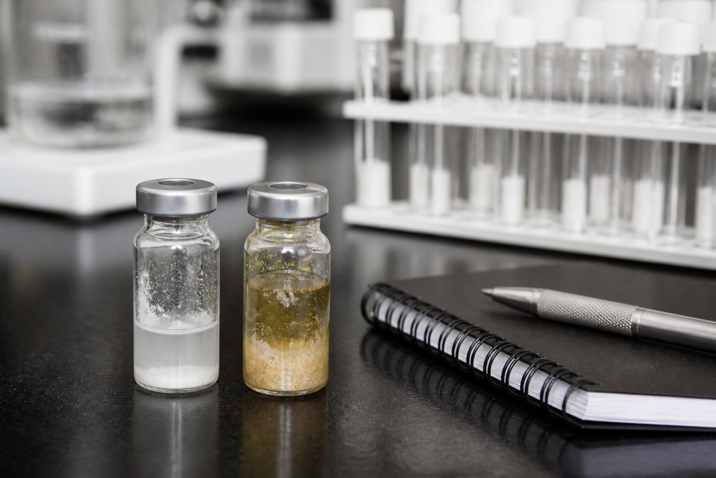
Fresh lyophilized peptides appear as consistent powder or cake, while reconstituted peptides in solution should be clear and colorless (unless the peptide contains aromatic amino acids like tryptophan or tyrosine).
Discoloration and Cloudiness
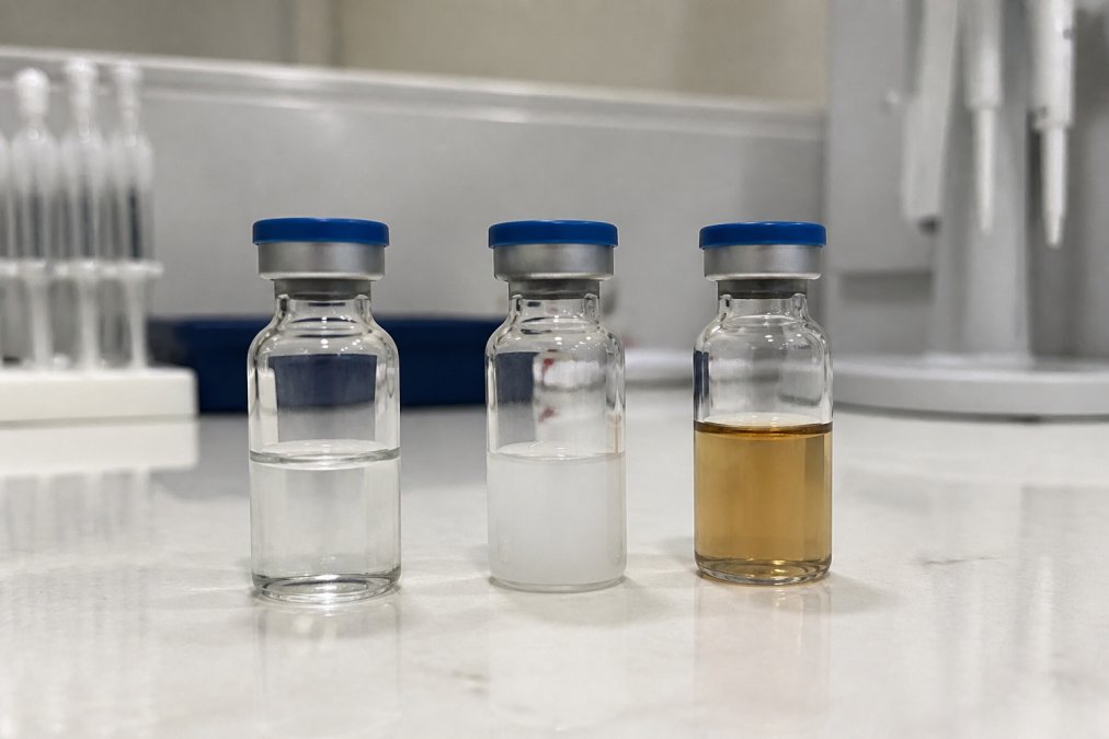
Discoloration signals chemical instability. Yellow, brown, or amber-tinted solutions indicate oxidation or covalent modifications where amino acid residues have reacted with oxygen or reactive species in storage.
Cloudiness indicates aggregation, individual peptide molecules clump together and precipitate out of solution, creating visible particles. This reduces the effective concentration of monomeric peptide and introduces unwanted particulates into assays. Storage conditions have been compromised through temperature fluctuations, light exposure, or contamination.
Particulate Matter and Precipitation
Visible particles represent advanced degradation from aggregated peptide molecules, microbial growth, or crystallized byproducts. Precipitation occurs when dissolved peptides can no longer remain in solution due to pH shifts, temperature changes, or molecular structure alterations from deamidation or proteolytic degradation.
Particulate matter clogs analytical instruments, interferes with spectrophotometric measurements, and compromises reproducibility.
Chemical Pathways Leading to Peptide Instability
Three primary mechanisms drive peptide instability: oxidation, hydrolysis, and deamidation. Understanding these pathways helps you recognize what's happening to your samples.
Oxidation and Amino Acid Residue Modification
Oxidation is the most common form of chemical instability. Exposure to oxygen attacks vulnerable amino acid residues, particularly methionine, tryptophan, and tyrosine.
Methionine oxidation is especially frequent. The sulfur atom in methionine's side chain converts to methionine sulfoxide, changing the peptide's charge, hydrophobicity, and conformation. A single oxidized residue shifts the entire peptide's behavior in your assay.
Tryptophan and tyrosine oxidation produces discoloration and alters binding affinity, enzymatic activity, and cellular uptake. Oxidation accelerates in the presence of light, heat, and metal ions, which is why amber vials, blocking ultraviolet and visible light, effectively slow degradation.
Hydrolysis and Sequence Integrity Loss
Hydrolysis breaks the peptide backbone itself. Water molecules attack peptide bonds, cleaving the sequence into smaller fragments. This process accelerates under acidic conditions, high temperatures, or in the presence of enzymes.
Hydrolysis produces a mixture of truncated fragments with different properties, lower molecular weight, altered charge, and reduced bioactivity. Your experiments show inconsistent results because you're no longer working with a pure compound. Early-stage hydrolysis isn't visually obvious, making analytical methods like mass spectrometry essential for confirmation.
Deamidation and Covalent Modification
Deamidation removes the amide group on asparagine or glutamine residues, converting them to aspartate or glutamate. This shifts charge and structure, often triggering aggregation of the entire peptide.
Deamidation accelerates at elevated temperature and alkaline pH. Once one or two residues undergo deamidation, the peptide's solubility decreases and it clumps with other partially degraded molecules, causing sudden cloudiness after weeks of room-temperature storage.
Peptide Storage Best Practices to Prevent Degradation
Prevention is the best management strategy. Storage conditions directly control the rate at which chemical pathways activate.
Temperature Control and Storage Conditions
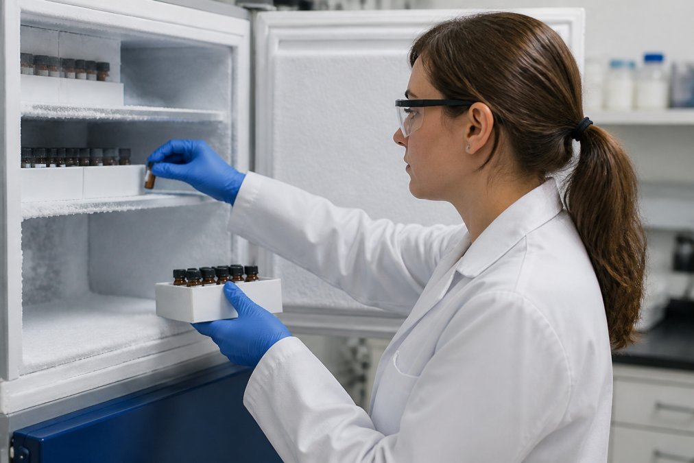
Temperature is the single most important variable in peptide stability. Chemical reactions accelerate exponentially with heat. Lyophilized peptides stored at -20°C or colder remain stable for years (peer-reviewed research). Room-temperature storage is acceptable only for days to a week.
Reconstituted peptides stored at 4°C remain usable for days to weeks; at room temperature, degradation may occur within hours or days (peer-reviewed research). At -20°C or -80°C, reconstituted peptides remain stable for months.
Minimize freeze-thaw cycles by aliquoting peptides into small portions before freezing. Use only what you need in a single experiment, then discard the remainder rather than refreezing.
Buffer Solutions and pH Sensitivity
The buffer solution controls pH, osmolarity, and solubility. Most peptides are supplied in optimized buffers, typically phosphate-based or acetate-based, and are most stable in the neutral to slightly acidic range (pH 4-7) (peer-reviewed research).
Use the buffer recommended by the supplier. A pH shift of even 0.5 units significantly accelerates deamidation or hydrolysis. For long-term storage, some peptides benefit from cryoprotectants like glycerol or DMSO, though these additives can interfere with certain assays.
Peptide Shelf Life After Reconstitution: What You Need to Know
Reconstitution marks the beginning of a stability window. Once the peptide is in solution, degradation accelerates significantly compared to the dry state.
A typical reconstituted peptide remains stable for 1-2 weeks at 4°C and several months at -20°C, depending on sequence, buffer composition, storage temperature, and handling. Plan experiments around reconstitution by creating small aliquots and freezing them individually.
Keep detailed records of reconstitution dates and usage. This historical data becomes invaluable if experimental results become inconsistent, you can correlate performance changes with material age.
Benefits of Amber Vials for Peptides and Long-Term Stability
Amber vials filter out ultraviolet and visible light, preventing photochemical reactions that accelerate oxidation. Light-driven oxidation is particularly problematic for peptides containing aromatic amino acids (tryptophan, tyrosine, phenylalanine) or sulfur-containing residues (methionine, cysteine).
Amber vials reduce light reaching the peptide by roughly 90% compared to clear glass, extending usable shelf life significantly. A peptide in an amber vial at 4°C may remain stable for weeks, while the same peptide in clear glass might degrade within days.
For long-term storage, amber vials are essential. Always request amber vials from suppliers and use them when transferring peptides to new containers.
Analytical Methods for Detecting Peptide Degradation
Visual inspection catches obvious degradation, but many forms of chemical instability are invisible. Analytical methods provide objective confirmation of peptide integrity.
Mass Spectrometry and HPLC Analysis
Mass spectrometry (MS) is the gold standard for detecting degradation by measuring molecular weight and revealing whether the peptide backbone has been cleaved or modified. High-performance liquid chromatography (HPLC) separates peptide molecules by chemical properties, showing whether your sample is a single pure compound or a mixture of degradation products.
Together, MS and HPLC provide a complete picture of peptide integrity. The downside is cost and accessibility, many labs lack these instruments in-house. Suppliers provide certificates of analysis documenting purity and integrity at manufacture.
BlueWaveCompound provides third-party lab-tested peptides with certificates of analysis for every batch, giving you documented assurance that material meets high-purity standards before you start experiments.
Practical DIY Testing Methods for Researchers
Visual inspection reveals cloudiness, discoloration, or visible particles indicating degradation.
Osmolarity testing detects aggregation through osmotic pressure changes.
pH testing confirms buffer hasn't drifted from expected range.
Protein concentration assays like Bradford or BCA detect peptide loss due to aggregation or precipitation.
Biological activity assays test whether the peptide binds receptors, activates enzymes, or produces cellular responses, the most relevant test for your research.
These methods provide quick, cost-effective screening to identify compromised samples before investing time in experiments.
Microbial Contamination as a Degradation Factor
Microbial contamination accelerates degradation dramatically. Bacteria and fungi secrete proteolytic enzymes that cleave peptide bonds, breaking down sequences into smaller fragments. Contamination also alters pH, consumes dissolved oxygen, or generates reactive chemical species.
Contamination is most likely in reconstituted peptides stored at room temperature or refrigerated. Prevention is far easier than remediation: use sterile technique when reconstituting, filter through 0.22-micron filters, and store at -20°C or colder. If you suspect contamination, discard the material.
Degradation vs. Impurity: Understanding the Difference
Impurity refers to non-peptide substances: salts, residual solvents, or synthesis byproducts. Impurities reduce effective peptide concentration but don't represent degradation. A 95% pure sample still contains intact, functional peptide molecules.
Degradation refers to chemical breakdown of the peptide itself, oxidation, backbone cleavage, aggregation, or modifications that alter structure or function. Degraded peptides are unsuitable regardless of purity.
Certificates of analysis report purity (percentage of target peptide) but may also include degradation products or modifications. High purity doesn't guarantee the peptide is intact, it only indicates what percentage is the target molecule, not whether that molecule is chemically unchanged.
Conclusion
Recognizing signs of degraded research peptides, from visual cues like discoloration to chemical mechanisms driving instability, is essential for maintaining experimental reproducibility.
Proper storage at low temperatures, protection from light using amber vials, and careful handling during reconstitution prevent most degradation. Analytical methods like mass spectrometry and HPLC provide definitive answers; practical DIY tests offer quick screening.
BlueWaveCompound delivers research-grade peptides with third-party lab testing and certificates of analysis for every batch, so you know exactly what you're working with before experiments begin. This quality assurance reduces the risk of experimental failure due to compromised materials and gives you confidence that your results reflect your science, not your reagents.
Frequently Asked Questions
How do I tell if my research peptides are degraded?
Degraded research peptides often show visual changes: cloudiness, discoloration, or particulate matter in solution. Lyophilized peptides may appear clumped or discolored. Chemically, degradation manifests through loss of secondary structure, aggregation, and reduced bioavailability. The most reliable method is analytical testing, HPLC analysis and mass spectrometry can confirm degradation by measuring changes in molecular weight and purity levels. If visual signs are present or experimental results show unusual noise, your peptides likely need replacement.
What environmental factors accelerate peptide breakdown?
Temperature fluctuations, exposure to light, humidity, and improper pH all accelerate peptide degradation. Oxidation occurs when peptides contact oxygen, particularly affecting methionine and tryptophan residues. Hydrolysis increases at higher temperatures and non-neutral pH levels. Microbial growth accelerates in reconstituted peptides stored above 4°C. Proteolytic degradation can occur if peptides contact contaminated surfaces. Storing lyophilized peptides at -20°C or colder, in amber vials, with desiccant, and in low-humidity conditions significantly extends shelf life.
How long do research peptides remain stable after reconstitution?
Reconstituted peptides typically maintain stability for 2-7 days when stored at 4°C in sterile buffer solutions, depending on the peptide sequence and buffer composition. Peptides with hydrophobic amino acid residues or prone to aggregation degrade faster. Adding preservatives or storing at -20°C extends the stability window to several weeks. Always note the reconstitution date and monitor for visual changes. Stability varies by peptide structure, so refer to your Certificate of Analysis or contact your supplier for sequence-specific stability data.
Why should I use amber vials for peptide storage?
Amber vials protect peptides from photodegradation by blocking ultraviolet and visible light wavelengths that trigger oxidation and deamidation. This protection is especially critical for lyophilized peptides and long-term storage. Amber vials reduce covalent modifications caused by light exposure, maintain amino acid residue integrity, and preserve bioavailability over extended shelf life. Combined with proper temperature control and desiccant packs, amber vials significantly reduce degradation rates compared to clear vials, making them standard in research-grade peptide storage protocols.
This article was written using GrandRanker